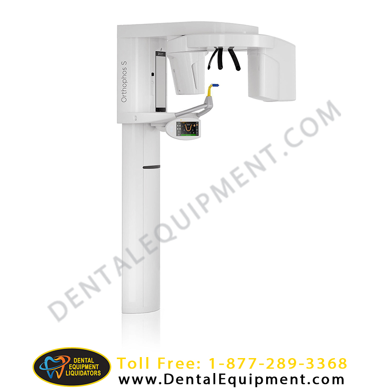
Sirona Digital Panoramic Orthophos S 2D X-ray *Used*
Stock: 3
Warranty: 1 Year Parts, 90 Days Labor
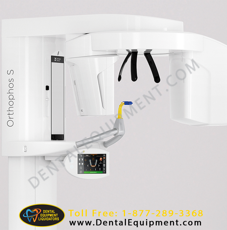
The high-quality 2D X-ray device with a comprehensive range of services for every practice is a reliable partner and optimized for everyday tasks. Its CsI Plus sensor with autofocus function ensures clear images, even in anatomically difficult cases. The automatic patient positioning together with the patented occlusal bite block enables an easy and timesaving patient positioning.
Selectable volume sizes
From Ø 5 cm x 5.5 cm to 8 cm x 8 cm or optional up to 11 cm x 10 cm
Easy to Use. Precise Patient Positioning
For most practices, they use an X-ray device with two main goals in mind: Capture the best possible image to support an accurate and precise diagnosis and ensure that the patient is comfortable during the process. The Orthophos S offers unique, patented solutions to help support both of these goals with:
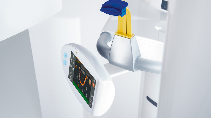
- The EasyPad and its intuitive user interface
- Automatic positioning aids such as the patented occlusal bite block and the 3-point head fixation
- Integrated temple width measurement
TECHNICAL DATA
- Nominal voltage: 200 – 240 V
- Permissible fluctuation: ± 10%
- Permissible drop under load: 10%
- Nominal current: 12A
- Nominal power output: 2 kW at 90 kV/12 mA with any radiation time
- Nominal frequency: 50 Hz / 60 Hz
- Mains resistance: Max. 0.8 ohms
- Main building fuse: 25 A slow-blow (16 A for single line)
- Power consumption: 2 kVA
- Power output of tube assembly: 69 kV / 16 mA = 1104 W with any radiation time
- Tube voltage: 60 – 90 kV (for 90 kV max. 12 mA)
- Tube current: 3 – 16 mA (for 16 mA max. 69 kV)
- Maximum setting range: 60 kV / 3 mA to 90 kV / 12 mA
- High-voltage waveform: High-frequency multipulse Residual ripple ≤ 4 kV
- High voltage generation frequency: 40 – 120 kHz
- Image acquisition scale: For P1, normal mandibular arch (slice center) approx. 1:1.25, i.e. the acquired image is magnified by approx. 24 % on average compared to reality.
- Exposure time for a cephalometric image: Max. 14.9 s
- Image acquisition scale for a cephalometric image: Approx. 1:1.1, i.e. the acquired image is magnified by approx. 10% on average compared to reality.
- Total filtration of X-ray tube assembly: > 2.5 mm AI / 90 IEC 60522 0.3 mm Cu for volume exposures 1 mm Cu for volume exposures in Low Dose mode
- Focal spot size as specified in IEC 60336, measured in the central X-ray beam: 0.5